The Optic Nerve Endings Are Located Within the
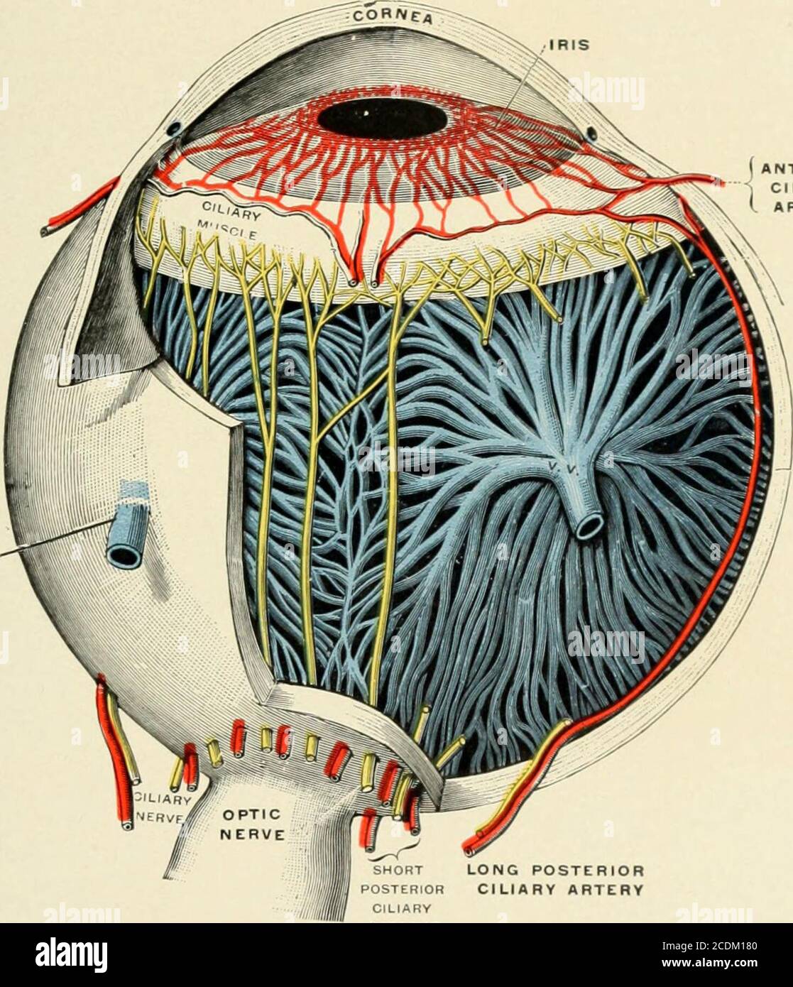
The optic nerve begins at the optic disk a structure that is 15 mm 006 inch in diameter and is located at the back of the eye. The relative excess optic nerve is.

Does The Inside Of The Body Have Nerve Endings Quora
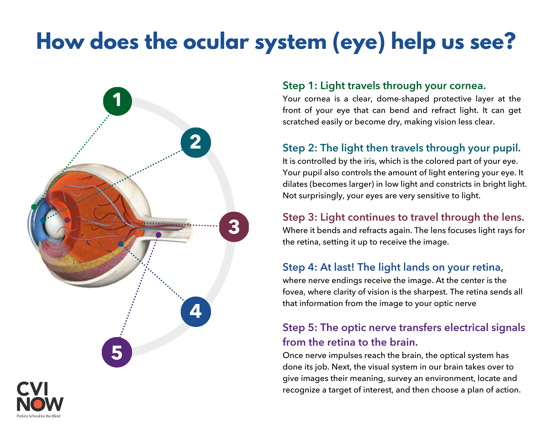
They are essentially little nerves with specialized endings called receptors which catch light particles called photons and convert the.
. Her pupils are bilaterally dilated and slow to react. The optic nerve endings are located within the. Fovea centralis macula lutea.
The optic nerve head is the most anterior component of the optic nerve and corresponds to the 1 mm segment that is located within the eyeball ie. 1 The straight-line distance from the back of the globe to the optic canal is much less the exact amount depending on individual orbital depth. The optic nerve senses the incoming light and image displayed on the retina.
Of the risk for airway problems. We call these cells by a general term known as photoreceptors. These cells release neurotransmitters onto a bipolar.
The optic nerve is a nerve made up of a bundle of Ganglionic cells located in the back of the eye which connects the eye to the brainIts function is to carry visual information in the form of electrical impulses from the retina to the areas of the brain which deal with vision allowing you to see these impulses as images in your head as your brain processes. Irrigate her eyes with water to prevent mucosal drying. A 39-year-old female experienced a severe closed head injury.
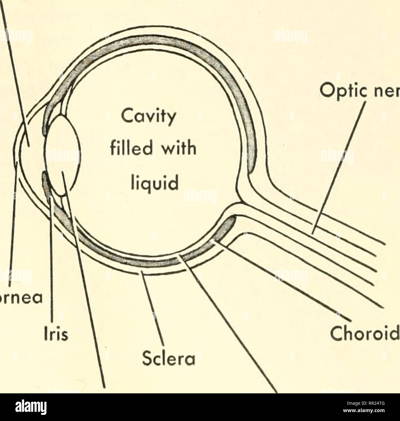
The optic nerve is located in the back of the eye. Asked Aug 29 2020 in Biology. The median eminence is a collection of nerve endings from neurosecretory cells that run the length of the pituitary stalk to the pituitary gland.
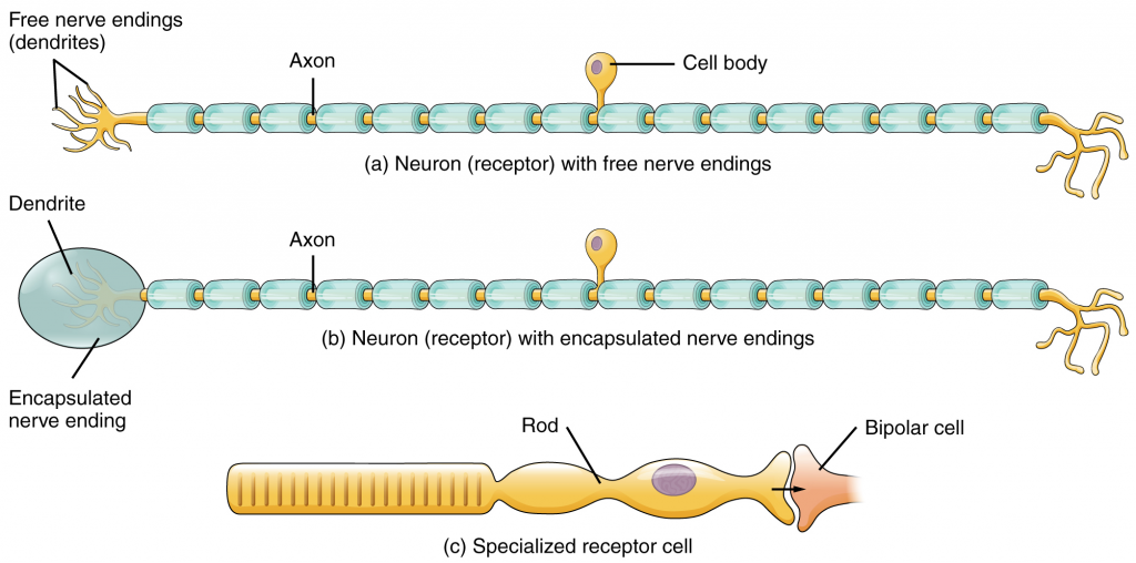
Photoreceptors in the eyes such as rod cells are examples of c specialized receptor cells. The relative excess optic nerve is. Asked Sep 17 2020 in Psychology by Hristo.
The optic nerve which acts like a cable connecting the eye with the brain actually is more like brain tissue than it is nerve tissue. The optic nerve also known as cranial nerve II is a continuation of the axons of the ganglion cells in the retina. Bleeding from soft-tissue injuries to the face is MOST effectively controlled with.
It is the second of several pairs of cranial nerves. Sharpness of image is the _____. The MOST significant complication associated with facial injuries is.
The optic nerve feeds in the posterior surface of the eye and carries all visual information from the retina ultimately to the primary orbital cortex which lies within the depths of the calcarine sulcus on the occipital lobe of the brain. The optic nerve also works in sync with the oculomotor nerve to change pupil size. The optic nerve also known as the second cranial nerve cranial nerve II or simply CN II is a paired cranial nerve that transmits visual information from the retina to the brain.
Asked Sep 28 2020 in Health Professions by styla. The optic nerve then transmits this image into the cerebral cortex. There are approximately 11 million nerve cells in each optic nerve.
Historically it was thought to be a raised entity protruding from the retinal surface and by extension was referred to as a papilla hence the term papilloedema. The rods and cones located in the retina are highly specialized neurons that convert information provided by light into electrical signals that are conducted to our brain. Optic Nerve CN II The function of the optic nerve is purely sensory in the eyes.
The fovea centralis is the region of the retina with a high concentration of cone cells colour detecting cells. The optic nerve endings are located within the. Asked Sep 28 2020 in Health Professions by styla.
Sensory neurons can have either a free nerve endings or b encapsulated endings. The optic nerve as well as the central artery and vein that. ____________ are sensory nerve endings found exclusively in the penis and clitoris.
It is located within a patch of cells posterior to the center of the lens called the _____. During inflammation as edema increases more pressure is exerted on nerve endings leading to increased. Free nerve endings tactile corpuscles and lamellated corpuscles are types of _____ receptors.
Abnormal variations in pupil size and reaction would MOST likely be observed in a patient with. Facial injuries should be identified and treated as soon as possible because. Other adjacent structures include the mammillary bodies and the optic chiasm.
The optic nerve endings are located within the. It extends from the optic disc to the optic chiasma. Emergency-medical-services _____ are sensory nerve endings found exclusively in the penis and clitoris.
AskedSep 28 2020in Health Professionsby styla. Sensory neurons can have either a free nerve endings or b encapsulated endings. During inflammation as edema increases more pressure is exerted on nerve endings leading to increased.
The optic nerve endings are located within the. In addition to managing problems with airway breathing and circulation you should. It is also called the second cranial nerve or cranial nerve II.
Direct pressure using dry sterile dressings. There is an approximately 1-mm component of optic nerve within the intrascleral part of the globe and approximately a 30-mm length of optic nerve from the globe to the optic canal. The optic nerve endings are located within the.
The skin and underlying tissues of the face. The optic nerve is located in the back of the eye. The optic nerve endings are located within the.
She is unresponsive with her eyes slightly open. Damage to the eyes. The optic nerve connects the eyes to several structures located within the diencephalon.
In humans the optic nerve is derived from optic stalks during the seventh week of development and is composed of retinal ganglion cell axons and glial cells. The optic disk forms from the convergence of ganglion cell output fibres called axons as they pass out of the eye.
A Treatise On Diseases Of The Eye Of Ranter 3 Terminal Nerve Fiberentering Epitiielial Layer 4 Nerve Ending Gutmann Nerve Trunks At The Nodes Of Llanvier Pass To Bowmmns Membrane Which They

197 Nerve Ending Illustrations Clip Art Istock

Week Two Lab Flashcards Quizlet

How Many Nerve Endings Are There In The Human Nose Quora
13 10 Sensory Perception Fundamentals Of Anatomy And Physiology

5 Histology Of Free Nerve Endings In Muscle Photographs Of Download Scientific Diagram
Which Part Of The Body Has The Greatest Concentration Of Nerve Ending To Make It Most Sensitive Quora

Pathway Of Normal Pain Perception Noxious Stimuli Activate Free Nerve Download Scientific Diagram

Nerve Endings Voetverzorging

6 Ultrastructure Of Free Nerve Endings A Reconstruction Of A Free Download Scientific Diagram

Biology Special Senses Lec 18 Flashcards Quizlet

Pin On Brain Anatomy And Function

197 Nerve Ending Illustrations Clip Art Istock

Muscle Spindle 3 4mm Extrafusal Intrafusal Muscle Fibers Alpha And Gamma Motor Neurons Sensory Axons And Human Heart Anatomy Motor Neuron Medical Anatomy

Chapter 10 Somatic Senses Ppt Download

7 Membrane Receptors Of A Nociceptive Nerve Ending Of Particular Download Scientific Diagram
How The Eyes Work Perkins School For The Blind
Adventures With Animals And Plants Biology 2 8o Why Liv Mg Receptors There Are Taste Buds On The Tongue And Special Nerve Endings For Smelhng In The Nose Scattered In The Skin

Biology Special Senses Lec 18 Flashcards Quizlet


Comments
Post a Comment